Coronal Plane Mri Spine

Mri Plane Cross Sections
my-ms.org

3d T2 Space Versus T2 Fse Or T2 Gradient Recalled Echo Which Is The Best Sequence American Journal Of Neuroradiology
www.ajnr.org
Spinal Sonography And Applications Of Ultrasound For Central Neuraxial Blocks Nysora
www.nysora.com

Magnetic Resonance Imaging Mri Scan Cervical Stock Photo Edit Now 721009741
www.shutterstock.com

Cureus Radiologic Evaluation Of Lumbar Spinal Stenosis The Integration Of Sagittal And Axial Views In Decision Making For Minimally Invasive Surgical Procedures
www.cureus.com
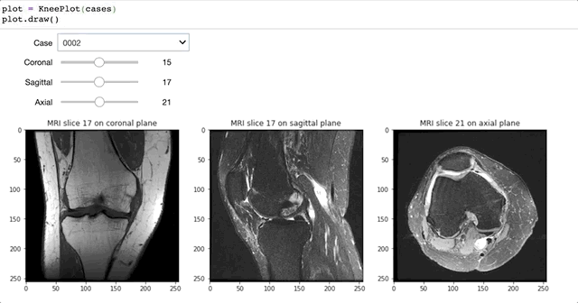
Analysis Of The Mrnet Knee Mri Dataset Towards Data Science
towardsdatascience.com
Tests in neurology and neurosurgery.
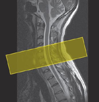
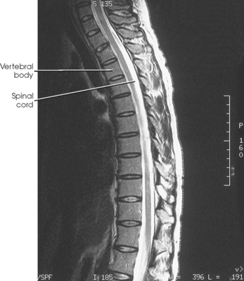
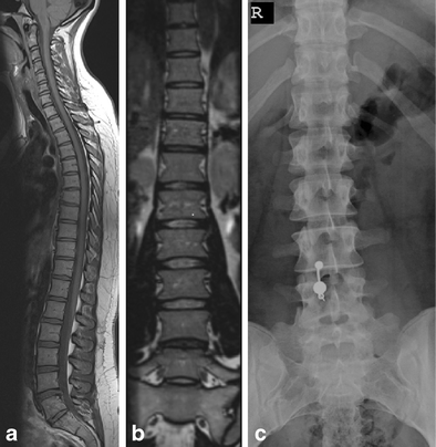
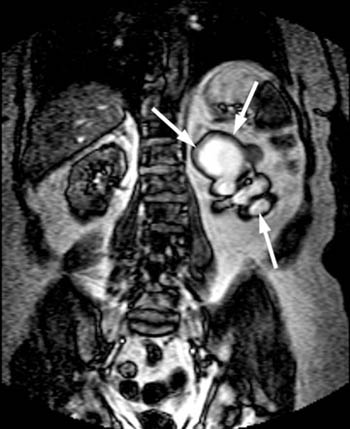
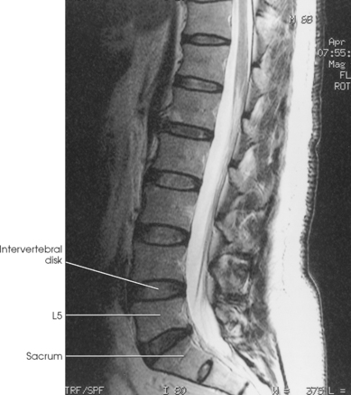
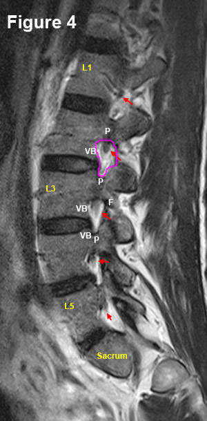
Coronal plane mri spine. 10a the scout coronal plane image shows a complex left adnexal mass in a post menopausal woman arrows. This axial t2 weighted image was part of a cervical spine mri examination. An unsuspected solid nodule arrow is present.
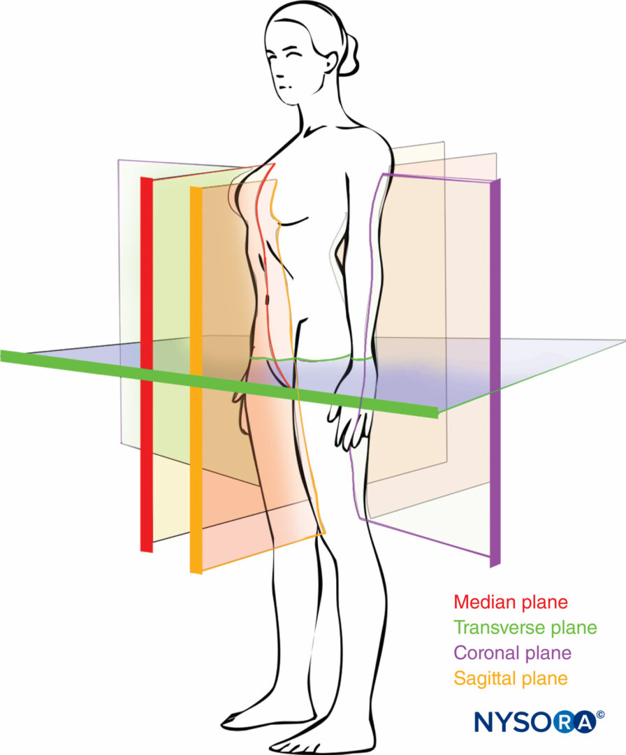
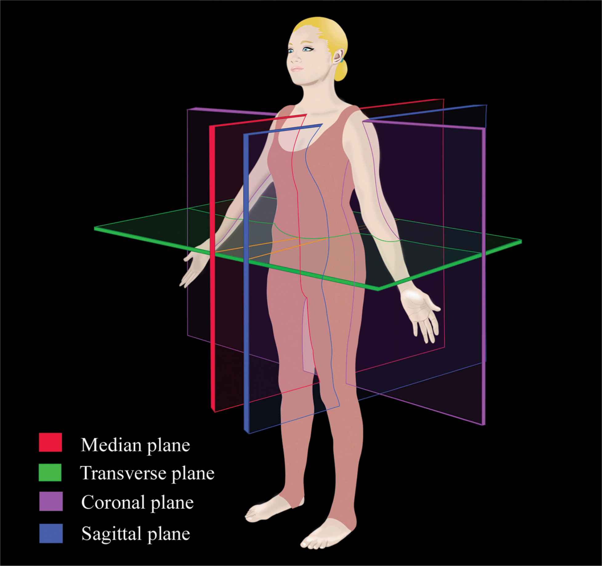
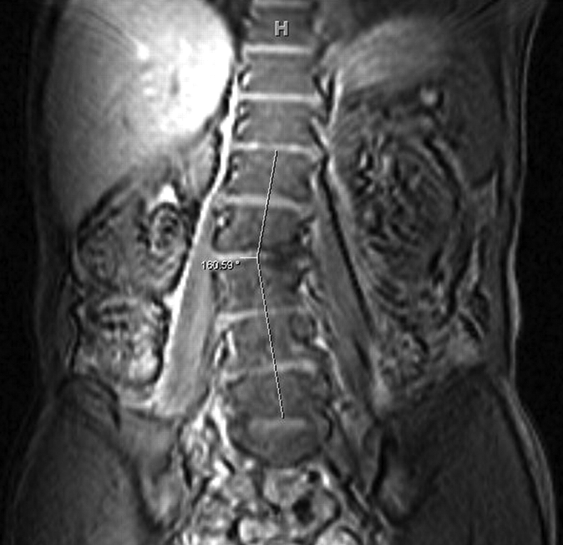
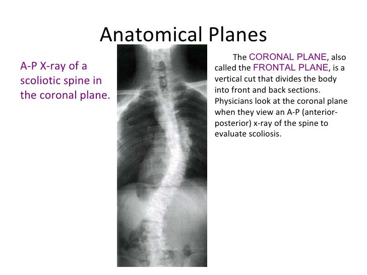
It measures whether or not the upper spine is located over the midline normal or off to one side. Prospectively we studied 32 patients average age 148 years with idiopathic scoliosis mean thoracic cobb angle 33 degrees on radiograph and 18. For a human the mid coronal plane would transect a standing body into two halves front and back or anterior and posterior in an imaginary line that cuts through both shoulders.
The coronal plane is an example of a longitudinal plane because it is perpendicular to the transverse plane. To assess coronal balance a vertical plumb line is drawn downwards from the mid point of the c7 vertebral body exactly in the same way as for the assessment of sagittal balance 1. The mri may scan your whole spine or just a part of it.
Axial sagittal and coronal see the example image below. Sagittal coronal and transverse slices anatomy of the lumbar vertebrae cross sectional imaging on t1 t2 and 3d mr. Supine mri does not show deformity well and in particular subluxations in the coronal plane.
Coronal balance is one of the features that needs to be assessed on long spine radiographs obtained for spinal deformity particularly scoliosis. Richter j tschoeke sk gulow j eichfeld u wojan m von salis soglio g heyde ce patient safety in surgery 2011. The coronal plane from the mobi view mri scan shows the open i therapeutic options to prevent recurrence of an aggressive aneurysmatic bone cyst of the cervical spine of a 16 year old boy a case report.
This type of lesion could represent a malignancy of arising from the left ovary and warrants further evaluation. Imaging in spine surgery 2017. Unfortunately many patients undergoing spinal surgery may only have supine mri of their spine.
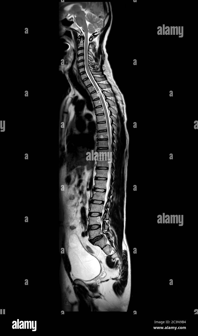
Its generally safe and painless. The coronal plane is often the most useful for evaluating bony anomalies spondylolysis or degeneration of the discs and facet joints. Unlike x rays and computerized tomography ct scans it doesnt use damaging radiation.
Mri provides exquisite detail of brain spinal cord and vascular anatomy and has the advantage of being able to visualize anatomy in all three planes.

Mri Of Cervical Spine Use Technique Myelogram On Coronal Plane For Diagnostic Spinal Cord Compression Stock Photo Download Image Now Istock
www.istockphoto.com

Spinal Cord Mri Protocols Planning And Indications Multiple Sclerosis Spine Mri Protocol
mrimaster.com
Http Pdf Posterng Netkey At Download Index Php Module Get Pdf By Id Poster Id 125274

Mri T2 Weighted Coronal Plane Image Idiopathic Scoliosis Patient 26 Download Scientific Diagram
www.researchgate.net

Racgp Making Sense Of Mri Of The Lumbar Spine
www.racgp.org.au
Spine Musculoskeletal Key
musculoskeletalkey.com
Mri Of Non Specific Low Back Pain And Or Lumbar Radiculopathy Do We Need T1 When Using A Sagittal T2 Weighted Dixon Sequence Springerlink
link.springer.com
Mri Of The Spines Of Children The Ispn Guide To Pediatric Neurosurgery
www.ispn.guide
Mri Protocols Techniques Used For Spondyloarthropathy 3 Tesla Mri Of The Spine
mriprotocol.blogspot.com

Mri Plane Mathematics
my-ms.org
%20mri%20planing%20and%20protocol%20of%20axial%20scans.JPG)
Spinal Cord Mri Protocols Planning And Indications Multiple Sclerosis Spine Mri Protocol
mrimaster.com

Anatomical Plane Wikipedia
en.wikipedia.org
Coronal Stir Sequence A Simple Adjustment To Routine Mri Protocol For Extra Spinal Sciatica And Sciatica Like Symptoms Springerlink
link.springer.com

Ct Scan Of Lumbar Spine
www.imaios.com
Cervical Spine Mri Planning Radtechonduty
www.radtechonduty.com

A Mri T2 Weighted Coronal Plane Shows A Degenerative Change In The Download Scientific Diagram
www.researchgate.net
Https Www Ejropen Com Article S2352 0477 18 30113 8 Pdf
Incidental Findings In Lumbar Spine Mri Their Prevalence And Potential Impact On Patient Management Egyptian Journal Of Radiology And Nuclear Medicine Full Text
ejrnm.springeropen.com

Approach To Mri Spine Learningneurology Com
learningneurology.com

Racgp Making Sense Of Mri Of The Lumbar Spine
www.racgp.org.au
Imaging Of Sacroiliitis Current Status Limitations And Pitfalls Tsoi Quantitative Imaging In Medicine And Surgery
qims.amegroups.com
Diagnostics Free Full Text Whole Body Mri With Diffusion Weighted Imaging In Bone Metastases A Narrative Review Html
www.mdpi.com

Spinal Cord Mri Protocols Planning And Indications Multiple Sclerosis Spine Mri Protocol
mrimaster.com
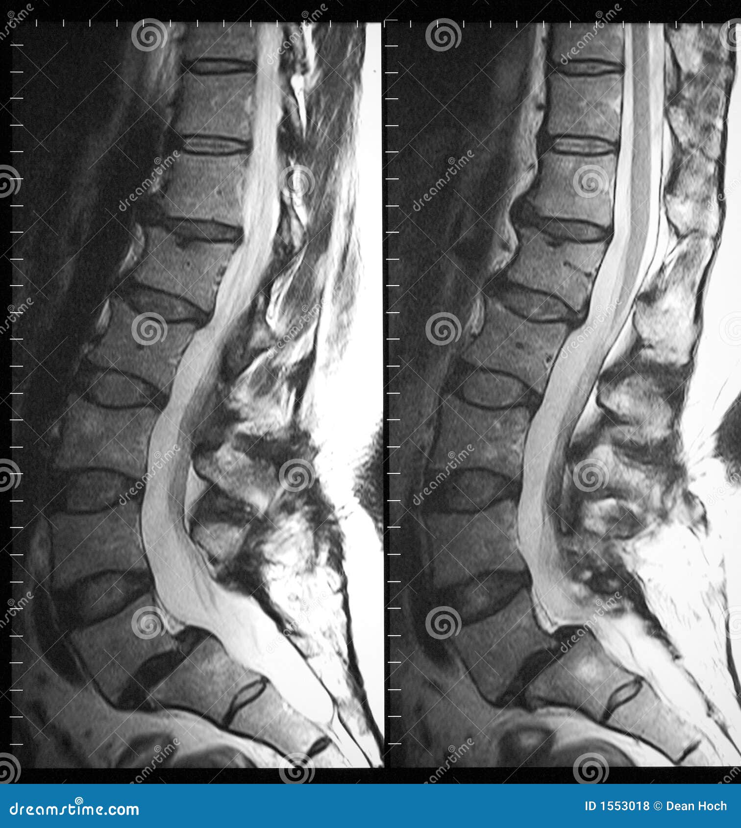
210 Lumbar Spine Mri Photos Free Royalty Free Stock Photos From Dreamstime
www.dreamstime.com

Lumbar Spine Anatomy On Mri Magnetic Resonance Imaging
www.imaios.com
Http Pdf Posterng Netkey At Download Index Php Module Get Pdf By Id Poster Id 125274
Disorders Of The Thoracic Spine Pre Operative Assessment And Surgical Management Chapter 10 Spine Disorders
www.cambridge.org
Mri Of The Spine And Bony Pelvis Radiology Key
radiologykey.com

Mri Protocols Pectoralis Muscle Mri Image Planes
mriprotocol.blogspot.com

Ivory Vertebra Imaging Findings In Different Diagnoses
www.scielo.br
The Radiology Assistant Ultrasound Of The Neonatal Spine
radiologyassistant.nl
Shutterstock Puzzlepix
shutterstock.puzzlepix.hu

Module 3 Abdominal Imaging
www.hitachimed.com
Vertebral Column Radiology Key
radiologykey.com

Module 5 Pelvis Imaging
www.hitachimed.com

Lumbar Lumbosacral Spine Mri Planning And Protocols Indications For Mri Lumbar Spine Scan
mrimaster.com
Primary Malignant Melanoma Of The Spinal Cord A Case Report
jcmtjournal.com
Read Your Mri Basic Education From A World Renowned Spine Expert Chirogeek Com
www.chirogeek.com
Radial Mr Screen Of The Sacroiliac Joints In The Lumbar Spine Of Patients With Chronic Lower Back Pain
clinmedjournals.org

Additional Merit Of Coronal Stir Imaging For Mr Imaging Of Lumbar Spine Gupta R Mittal P Mittal A Mittal K Gupta S Kaur R J Craniovert Jun Spine
www.jcvjs.com
Incidence Of Numerical Variants And Transitional Lumbosacral Vertebrae On Whole Spine Mri Springerlink
link.springer.com

Ct Scan Wikipedia
en.wikipedia.org

Magnetic Resonance Imaging Mri Scanning Principles Teachmeanatomy
teachmeanatomy.info

Anatomy Of Spine
www.slideshare.net
Spinal Sonography And Considerations For Ultrasound Guided Central Neuraxial Blockade Anesthesia Key
aneskey.com
Plos One Pelvic Belt Effects On Pelvic Morphometry Muscle Activity And Body Balance In Patients With Sacroiliac Joint Dysfunction
journals.plos.org
Sagittal Plane High Resolution Stock Photography And Images Alamy
www.alamy.com
Plos One Pelvic Belt Effects On Pelvic Morphometry Muscle Activity And Body Balance In Patients With Sacroiliac Joint Dysfunction
journals.plos.org
Incidental Findings On Musculoskeletal Mr Radsource
radsource.us

Spinal Cord Mri Protocols Planning And Indications Multiple Sclerosis Spine Mri Protocol
mrimaster.com
The Coronal Plane From The Mobi View Mri Scan Shows The Expansiveness Download Scientific Diagram
www.researchgate.net
Http Pdf Posterng Netkey At Download Index Php Module Get Pdf By Id Poster Id 125274

Mri Of The Dorsal Spine T2weighted Coronal Plane And T1 Weighted Download Scientific Diagram
www.researchgate.net

Adult Spinal Deformity Spine Orthobullets
www.orthobullets.com
Https Encrypted Tbn0 Gstatic Com Images Q Tbn 3aand9gctjwpwibk8ed58qepbsidmdyp3mp8hi9sawovkj0xzyo7cav9zn Usqp Cau
encrypted-tbn0.gstatic.com
Adult Lumbar Scoliosis Underreported On Lumbar Mr Scans American Journal Of Neuroradiology
www.ajnr.org
Vertebral Column Radiology Key
radiologykey.com

Additional Merit Of Coronal Stir Imaging For Mr Imaging Of Lumbar Spine Gupta R Mittal P Mittal A Mittal K Gupta S Kaur R J Craniovert Jun Spine
www.jcvjs.com

Lumbar Lumbosacral Spine Mri Planning And Protocols Indications For Mri Lumbar Spine Scan
mrimaster.com
Mri Of The Spines Of Children The Ispn Guide To Pediatric Neurosurgery
www.ispn.guide
Radiography Imaging Planes Radtechonduty
www.radtechonduty.com
Would An Mri Scan Of An Ls Spine Include An Si Joint Quora
www.quora.com
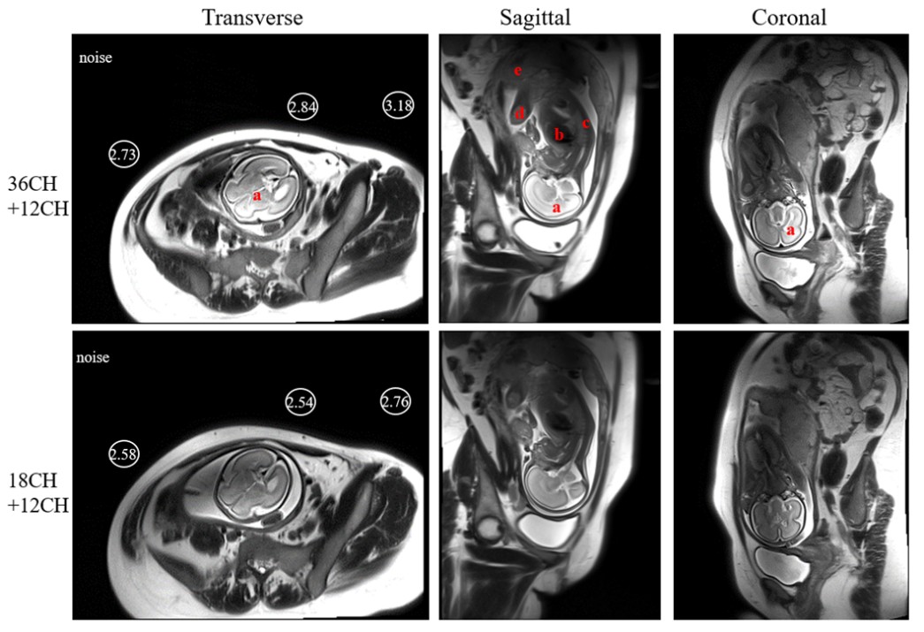
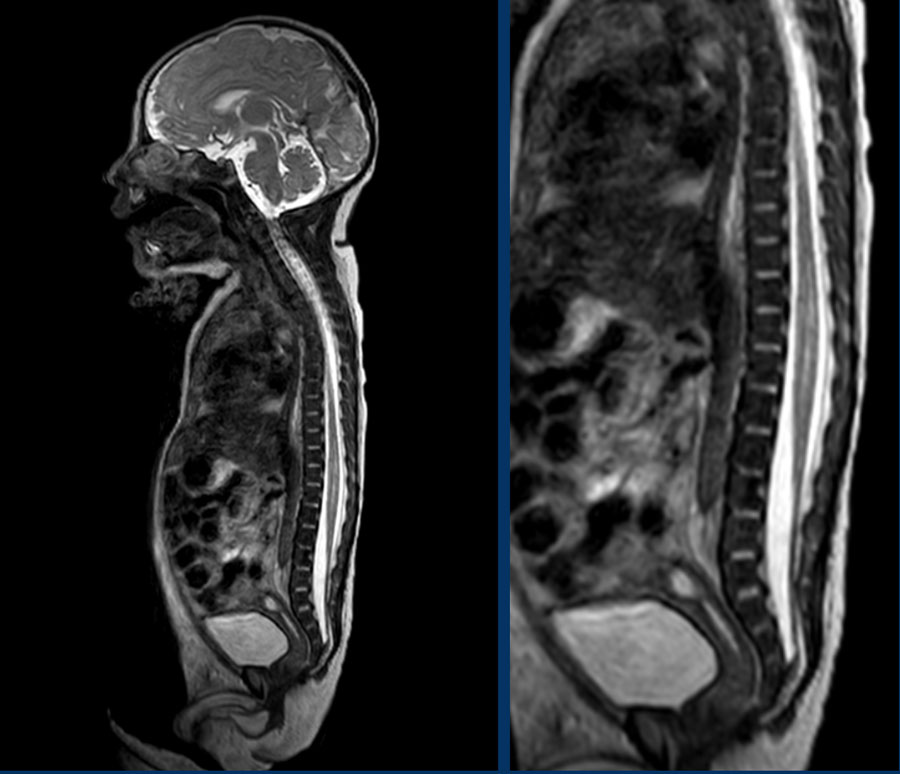
Dedicated 36 Channel Receive Array Breaks Through The Limitation Of Existing Mri For Fetal Shenzhen Institutes Of Advanced Technology
english.siat.cas.cn

Atlas Jhp Mri Brain Atlas
jhpmribrainatlas.rcc.uchicago.edu
Anatomy Of Spine
www.slideshare.net

Thoracic Spine Mri Planning Mri Thoracic Spine Protocols Indications For Mri Thoracic Spine Scan
mrimaster.com

Subdural Hematoma On Post Contrast Post Gadolinium T1 Mri In The Coronal Plane Subdural Hematoma Mri Healthcare Professionals
www.pinterest.com

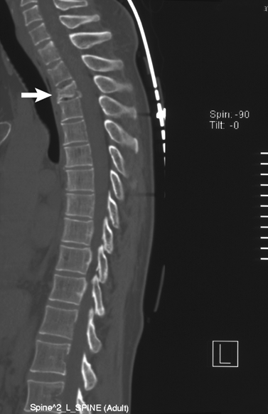
Ct Scan Of Lumbar Spine
www.imaios.com
The Thoracic Spine Radiology Key
radiologykey.com

Thoracic Spine Mri Planning Mri Thoracic Spine Protocols Indications For Mri Thoracic Spine Scan
mrimaster.com
How To Read The Normal Knee Mri Kenhub
www.kenhub.com
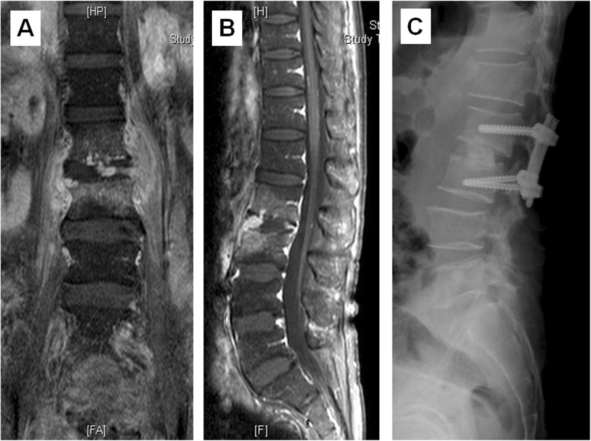
Comparison Of Two Stage Open Versus Percutaneous Pedicle Screw Fixation In Treating Pyogenic Spondylodiscitis Bmc Musculoskeletal Disorders Full Text
bmcmusculoskeletdisord.biomedcentral.com

Imaging The Cervical Thoracic And Lumbar Spine Radiology Key
radiologykey.com
Figure 2 From Unusual Variant Of Coronal Bladder Duplication Associated With Glans Diphallia A Case Report And Review Of The Literature Semantic Scholar
www.semanticscholar.org
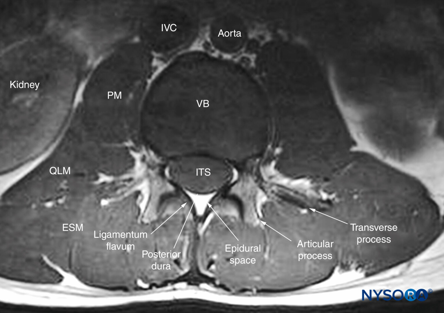
Spinal Sonography And Applications Of Ultrasound For Central Neuraxial Blocks Nysora
www.nysora.com
Https Encrypted Tbn0 Gstatic Com Images Q Tbn 3aand9gcr11pd R4xf1kinwwbjqnpxlc53mmbqlxqweggjrazknb544j2e Usqp Cau
encrypted-tbn0.gstatic.com

Medpix Case Left C5 C6 Disc Herniation With Radiculopathy
medpix.nlm.nih.gov
Https Encrypted Tbn0 Gstatic Com Images Q Tbn 3aand9gctjwpwibk8ed58qepbsidmdyp3mp8hi9sawovkj0xzyo7cav9zn Usqp Cau
encrypted-tbn0.gstatic.com

The Lumbar Spine Mri Taken In March 2010 A The Sagittal Plane Download Scientific Diagram
www.researchgate.net

Ct Scan Of Cervical Spine Stock Footage Video 100 Royalty Free 1021089349 Shutterstock
www.shutterstock.com

Adult Spinal Deformity Spine Orthobullets
www.orthobullets.com
Cervical Spine Mri Planning Radtechonduty
www.radtechonduty.com
Mri Plane Mathematics
my-ms.org

Lumbar Mri Part 1 Normal Imaging Appearance Of The Lumbar Spine Sciencedirect
www.sciencedirect.com

Mri Basics
casemed.case.edu
Figure 3 From Osteochondroma Of The Thoracic Spine And Scoliosis Semantic Scholar
www.semanticscholar.org

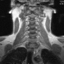
Cervical Spine Mri
www.imaios.com
Https Encrypted Tbn0 Gstatic Com Images Q Tbn 3aand9gcrcknj4aq4odfyvegratcu Xkdiunokryaovfh0viaty5mh5cvz Usqp Cau
encrypted-tbn0.gstatic.com

Abnormal Archives Sohrab Gollogly Md
sohrabgolloglymd.com
Normal Mri Of Thoracic Spinal Cord Stock Photo Alamy
www.alamy.com

Cross Sectional Study On Incidental Spinal Findings In Magnetic Resonance Imaging Lumbar Spine Of Patients With Low Back Pain
wajradiology.org

Evaluation Of Traumatic Spine By Magnetic Resonance Imaging And Its Correlation With Cliniconeurological Outcome Naik Br Sakalecha Ak Savagave Sg J Emerg Trauma Shock
www.onlinejets.org
Read Your Mri Basic Education From A World Renowned Spine Expert Chirogeek Com
www.chirogeek.com

Exemplary Mri Images Of A Pig Spine In A Sagittal A And Coronal B Download Scientific Diagram
www.researchgate.net
Understanding Medical Tests Mri Magnetic Resonance Imaging
www.coloradospineinstitute.com

Cross Sectional Study On Incidental Spinal Findings In Magnetic Resonance Imaging Lumbar Spine Of Patients With Low Back Pain
wajradiology.org
The Potential Risk Of Spinal Cord Injury From Pedicle Screw At The Apex Of Adolescent Idiopathic Thoracic Scoliosis Magnetic Resonance Imaging Evaluation Bmc Musculoskeletal Disorders Full Text
bmcmusculoskeletdisord.biomedcentral.com
Medical Imaging And Radiological Anatomy X Ray Ct Mri Kenhub
www.kenhub.com

